 Life & Style

Đà Nẵng aims to rank among Asia’s leading cities by 2075
1.
Dr Saiko Sugiyama*
Breast cancer is one of the most common cancers affecting women worldwide. While screening recommendations differ slightly between countries, they are all designed to balance early detection with safety and accuracy.
This article summarises current evidence-based screening guidance and explains why individualised screening – especially for younger women and those planning pregnancy – is important.
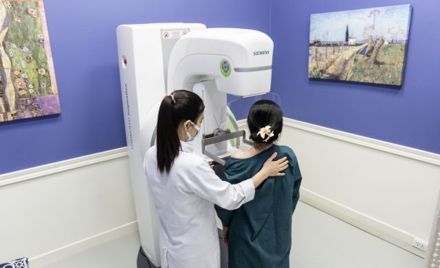
|
| 3D Mammography machine. — Photo courtesy of Family Medical Practice |
1) Recommended screening frequency
United States
Most major organisations recommend screening mammography starting at age 40 for women at average risk.
Depending on the guideline, screening is performed annually or every two years, continuing into the early 70s as long as the woman remains in good health.
United Kingdom
In the UK, the National Health Service (NHS) provides population-based screening every three years, generally between the ages of 50 and 71. Women outside this age range are encouraged to seek medical evaluation promptly if they notice symptoms.
Japan
In Japan, population-based breast cancer screening is centred on mammography for women aged 40 years and older, commonly performed every two years. Ultrasound is widely used in clinical practice but is not uniformly recommended for population screening.
Important note:
These age thresholds are based on public health efficiency. They do not imply that breast cancer does not occur in younger women.
2) Mammography and ultrasound: differences, advantages and limitations
Mammography
Advantages
Limitations
Breast ultrasound
Advantages
Limitations
Clinical perspective:
For average-risk women, mammography remains the foundation of screening. Ultrasound plays an important adjunctive or individualised role, particularly in younger women and during pregnancy.
3) Breast cancer risk in younger women and the role of early assessment
Although most screening guidelines begin at age 40 or later, breast cancer incidence begins to rise from the late 20s and 30s. Breast cancer is consistently among the most common cancers in women, and a meaningful proportion of cases occur before the age of 40.
Why diagnosis can be delayed in younger women
These factors are clinically important because childbearing age overlaps with the period when breast cancer risk begins to increase.
Pregnancy as a diagnostic challenge
As pregnancy progresses, breast tissue undergoes rapid physiological changes, including glandular proliferation and ductal development. These normal changes can make both physical examination and imaging interpretation more difficult, potentially delaying diagnosis of pre-existing disease.
Why early pregnancy (or preconception) matters
In early pregnancy, before significant breast enlargement occurs, abnormal findings are easier to detect.
From a clinical standpoint, early assessment allows:
Breast ultrasound is safe during pregnancy and does not pose a risk to the fetus, making it well suited for younger women and pregnant patients.
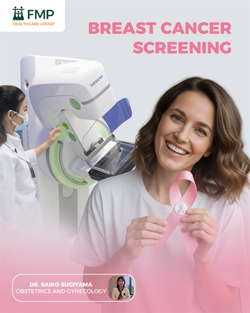
|
| Most major organisations recommend screening mammography starting at age 40 for women at average risk. — Photo courtesy of Family Medical Practice |
4) Breast cancer screening when trying to conceive or in very early pregnancy
Women who are planning pregnancy or are in very early pregnancy often postpone health checks. However, this period may actually represent a valuable opportunity to assess baseline breast health.
For women near screening age or with additional risk factors, completing appropriate breast evaluation before conception or in early pregnancy can be both practical and reassuring.
5) Hormonal contraception and hormone replacement therapy (HRT)
Hormonal contraception (including combined oral contraceptives)
Current evidence suggests a small increase in breast cancer risk during active or recent use, which gradually returns to baseline after discontinuation. This represents a population-level risk and does not mean most users will develop breast cancer.
Hormone replacement therapy
Systemic combined estrogen–progestin therapy has been associated with an increased breast cancer risk, particularly with longer duration of use.
Clinical implication:
Women using hormonal therapies should ensure they are up to date with age-appropriate screening and seek evaluation promptly if symptoms occur.
6) Family history: when does risk clearly increase?
Family history becomes clinically significant when certain patterns are present, particularly:
In such cases, individualised risk assessment and referral for specialist evaluation may be appropriate.
7) Other lifestyle-related risk factors
Alcohol
Alcohol consumption is a well-established risk factor for breast cancer. Risk increases with intake, and even low levels may contribute. From a prevention perspective, less alcohol is better.
Smoking
Smoking has also been associated with increased breast cancer risk, with evidence suggesting a dose–response relationship.
Key take-home messages
*Dr Saiko Sugiyama is a highly skilled obstetrician and gynaecology with international medical training and extensive clinical experience in women’s health. She graduated from medical school in Japan and has worked in both hospital and clinical settings, providing comprehensive care across obstetrics and gynecology.
Dr Sugiyama specialises in antenatal and postnatal care, routine gynaecological examinations, and the diagnosis and management of a wide range of women’s health conditions. She is known for her patient-centred approach and clear communication, supporting women through every stage of life.
FMP Healthcare Group operates medical centres in major cities including HCM City, Hà Nội and Đà Nẵng, offering consultations with international doctors, check-up centres and emergency ambulance services.
Visit FMP Thảo Điền in HCM City to consult with Dr Saiko Sugiyama.
To book an appointment, please call (028) 3744 2000 (FMP - Thảo Điền), or email d2.ob-gyn@vietnammedicalpractice.com.
Life & Style
 Life & Style
.jpg) Life & Style
Life & Style
 Life & Style
 Life & Style
 Life & Style
 Life & Style
 Life & Style
 Life & Style
 Life & Style
 Life & Style
 Life & Style
 Life & Style
 Life & Style
 Life & Style