 Life & Style

Dr Andres Sosa *
 |
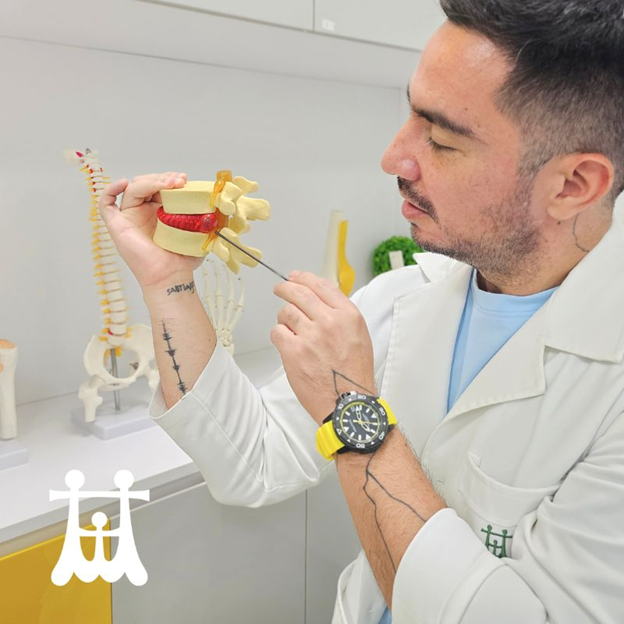
| Image-guided facet joint injections: minimally invasive relief for cervical and lumbar disc herniation pain. — Photo courtesy of Family Medical Practice |
In the bustling city of Hà Nội, where expatriates, diplomats and tourists often find themselves immersed in sedentary work, orthopedic issues like cervical and lumbar disc herniation pain can be particularly prevalent. As an orthopedic surgeon committed to the international community in the north of Việt Nam, offering effective solutions is crucial.
Understanding image-guided facet joint injections
Facet joints of the spine are small joints located between each vertebra. They play an important role in the stability and movement of the body. When these joints become inflamed or irritated they can contribute significantly to chronic neck or lower back pain. One of the most common causes of these inflammation is disc herniation. This is a condition in which the intervertebral disc, which acts as a cushion between the vertebrae in the spine, bulges or ruptures.
The procedure involves injecting a combination of a local anesthetic and a corticosteroid directly into the affected facet joints under the guidance of imaging techniques such as fluoroscopy or C-arm fluoroscopy. This precision ensures accurate placement of the needle, maximising the effectiveness of the injection.
Indications and patient selection
Image-guided facet joint injections are typically recommended for patients who have been diagnosed with facet joint syndrome through imaging studies like CT scans or MRI and who have not achieved sufficient relief from conservative management approaches such as oral analgesics and physical therapy.
Common indications include:
- Chronic neck or lower back pain that radiates into the arms or legs.
- Pain worsened by specific movements or positions, characteristic of facet joint involvement.
- Failure to respond adequately to conservative treatments.
Patient selection involves a thorough evaluation to confirm that the facet joints are indeed the source of the pain. This evaluation ensures that the injection will be both diagnostic and therapeutic, confirming the diagnosis while providing immediate pain relief.
Benefits of image-guided facet joint injections
1. Minimally invasive: compared to surgical interventions, image-guided facet joint injections are minimally invasive procedures performed on an outpatient basis. This minimises recovery time and reduces the risk of complications.
2. Precise targeting: the use of imaging guidance (fluoroscopy or C-arm) allows for precise targeting of the affected facet joints, ensuring that the medication is delivered directly to the source of pain.
3. Immediate and long-lasting relief: the injection provides immediate relief from pain due to the local anesthetic, while the corticosteroid reduces inflammation and can provide long-lasting pain relief ranging from weeks to months.
4. Complementary to physical therapy: facet joint injections are often used in conjunction with physical therapy. While injections provide relief, physical therapy helps to strengthen muscles, improve flexibility and correct postural imbalances, which can contribute to long-term pain relief and prevention of future episodes.
Procedure and recovery
Before the procedure, patients are typically advised to avoid eating or drinking for a few hours. The procedure itself involves:
- Preparation: The patient lies on an X-ray table and the skin over the injection site is cleaned and numbed with a local anesthetic.
- Injection: The physician uses fluoroscopy or C-arm fluoroscopy to guide the needle into the facet joint. Once the needle is correctly positioned, the medication is injected.
- Post-procedure: Patients are monitored briefly and then discharged with instructions. Mild soreness at the injection site is common and can be managed with ice and over-the-counter pain medications.
Considerations and risks
While generally safe, image-guided facet joint injections may carry some risks, including infection, bleeding, or allergic reactions to the medications used. These risks are minimised through proper sterile technique and careful patient selection.
Conclusion
In conclusion, image-guided facet joint injections represent a valuable tool in the management of cervical and lumbar disc herniation pain, particularly for patients who have not responded adequately to conservative treatments.
As an orthopedic surgeon in Hà Nội, offering these advanced treatments underscores my commitment to providing comprehensive care to my patients. By integrating image-guided facet joint injections with my expertise in orthopedic care, I continue to enhance patient outcomes and quality of life in my practice. Family Medical Practice
* Dr Andres Sosa is our orthopedic surgeon specialising in sports medicine and trauma. After his residency in orthopedics, he took a master’s in upper limb surgery at the University of Bologna (Italy), attended the sports medicine programme at Thomas Jefferson University in Philadelphia (USA) and obtained his second master’s in shoulder surgery with the University of Andalucía (Spain). Once in Việt Nam, he continues his surgical training with Arthrex ArthroLab (Singapore) focused on arthroscopic techniques for shoulder and knee injuries.
Dr Sosa joined FMP in 2018 and is responsible for all orthopedic and trauma cases. He is also a sports nutrition expert from Major University (Chile) and is fluent in English, Italian and Spanish.
Visit Family Medical Practice Hanoi 24/7 at 298I Kim Mã, Kim Mã Ward, Ba Đình District.
To book an appointment, please call us at (024).3843.0784 or via Whatsapp, Viber or Zalo on +84.944.43.1919 or email hanoi@vietnammedicalpractice.com.
FMP’s downtown location in Hồ Chí Minh is in Diamond Plaza, 34 Lê Duẩn Street, Bến Nghé Ward, District 1, and 95 Thảo Điền Street, District 2. Tel. (028) 3822 7848 or email hcmc@vietnammedicalpractice.com.


