 Society

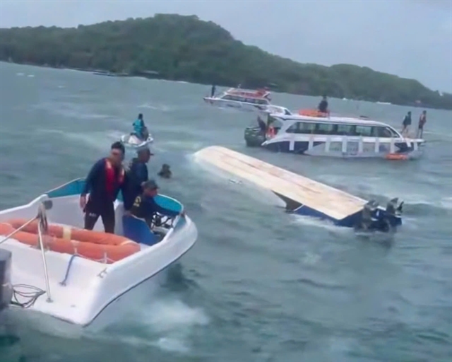
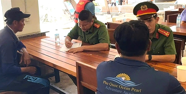
Repatriation and support efforts underway after deadly Phú Quốc capsizing
1.
 |
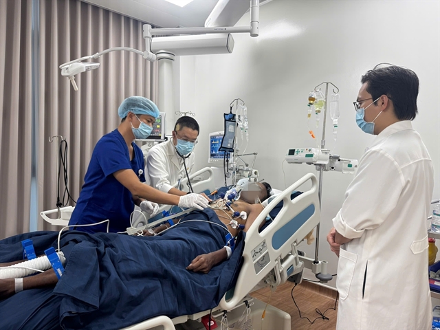
| Doctors perform the surgery for a 49-year-old woman, who had suffered a deep knife wound to her heel, at the National Hospital for Tropical Diseases in Hà Nội.— Photo courtesy of the National Hospital for Tropical Diseases |
HÀ NỘI — In a groundbreaking operation, doctors at the National Hospital for Tropical Diseases have successfully reconstructed nearly the entire Achilles tendon of a 49-year-old woman who suffered a deep knife wound to her heel, saving her from permanent disability and restoring up to 90 per cent of her mobility.
Dương Mạnh Chiến, consultant plastic and reconstructive surgeon at the hospital, said: “This is the first time our hospital has performed simultaneous reconstruction of tendon and skin using a vascularised ‘chimeric flap’ technique in a case of severe infection. Very few medical centres in Việt Nam are currently capable of carrying out such complex microsurgery.”
Initially, the patient’s wound was sutured at a local medical facility without detecting that the Achilles tendon had been severed. When her ability to walk failed to improve, she was diagnosed with a tendon rupture and underwent repair surgery at another hospital.
However, after the operation, the wound developed an abscess and severe necrotising infection, requiring a second procedure to clean the site. Despite this, the wound continued to deteriorate, discharging pus and failing to heal. The entire 10–12 cm length of the Achilles tendon had become necrotic and the surrounding soft tissue was destroyed, leaving the patient almost unable to move her foot.
According to Chiến, the surgical team had to proceed in two stages to treat the complex combination of deep infection and loss of both tendon and skin.
“In the first stage, we removed all necrotic tissue, drained the abscess and used a vacuum-assisted closure (VAC) system to control the infection,” he said. “Once the wound bed was clean, we performed microsurgical reconstruction using a ‘chimeric flap’, a composite graft taken from the thigh that includes both tendon and skin, nourished by a shared vascular pedicle, which was then connected to blood vessels in the heel.”
A key feature of the operation was the use of the fascia lata tendon — a strong, elastic structure from the thigh — to recreate the Achilles tendon. The harvested tendon was rolled and shaped to resemble the original and anchored to the heel bone. At the same time, a vascularised skin flap was transferred to cover the large tissue defect.
Both flaps were sustained through delicate microsurgical anastomosis performed under a high-powered operating microscope magnified up to 40 times, enabling precise handling of blood vessels thinner than a strand of hair.
Chiến said the procedure demanded not only advanced microsurgical skills but also sound clinical judgement, balancing infection control, identifying viable tissue and selecting graft material with closely matched biomechanical properties.
Following surgery, the patient has regained almost full walking ability — a remarkable recovery given that the Achilles tendon is essential for bearing body weight during movement.
Drawing lessons from the case, Chiến said tendon and soft-tissue injuries must be properly assessed from the outset. Simply stitching the skin while overlooking a tendon rupture could result in severe loss of function. Maintaining sterility after surgery was also critical to prevent infection and necrosis.
“If the Achilles tendon has become necrotic, the patient must be referred to a specialist reconstructive centre capable of restoring both structure and function,” he said.
He also urged the public to seek prompt medical attention at specialised hospitals if a wound fails to heal or shows signs of swelling, pain or discharge. For complex injuries of this nature, only centres with expertise in microsurgery and modern equipment can manage the condition effectively, preserving not only mobility but also the patient’s quality of life. — VNS
Society
 Society
 Society
 Society
 Society
 Society
 Society
 Society
 Society
 Society
 Society
 Society
.jpeg) Society
Society
 Society
 Society