 Society


Việt Nam, China strengthen exchanges between military academies
1.
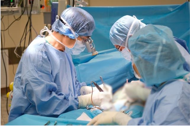 |
| Dr. Lương Ngọc Trung and his surgical team remove a lung tumor from a patient. — Photo courtesy of the hospital |
HCM CITY — A lung cancer patient, just days before surgery, was unexpectedly found to have a giant cerebral arteriovenous malformation that could rupture at any moment during the operation.
The story began more than a month ago when Đ.N.N. (58 years old in HCM City) experienced a persistent cough and sought a check-up at a major private hospital. The results revealed an abnormal white spot on her lung. The doctor recommended immediate surgery, followed by a biopsy to determine whether the tumor was benign or malignant.
Feeling anxious and uncertain, N. and her husband decided to seek a second opinion at FV Hospital.
Dr. Lương Ngọc Trung, head of the Thoracic, Vascular, Endovascular Surgery Department at FV Hospital, examined her and noted that imaging revealed a lung opacity of over 2cm. According to the Cancer Society guidelines, such a pulmonary nodule requires careful monitoring and appropriate management.
A CT-guided needle biopsy confirmed lung cancer.
However, during an MRI brain scan to check for metastasis, doctors discovered a giant cerebral arteriovenous malformation - an abnormal tangle of blood vessels in the brain - that could rupture at any time, posing a high risk during anesthesia for surgery.
To determine the optimal surgical approach, FV Hospital convened a multidisciplinary consultation with in-house and external cerebrovascular specialists, and also sought input from experts in Singapore via Thomson Medical Group.
The specialists agreed that the cerebral arteriovenous malformation did not require urgent intervention. While lung surgery carried a risk of complications, it was considered manageable if critical factors, such as blood pressure, were well controlled.
On the morning of July 31, after meticulous preparation, surgeons performed a minimally invasive thoracoscopic procedure through a 3cm incision. In three hours, the lung tumor was completely removed. Pathology results confirmed stage 1A lung cancer with no lymph node metastasis, indicating an excellent prognosis that required only regular follow-up and no additional treatment. The cerebral arteriovenous malformation caused no complications during surgery.
Seven days after the operation, the patient was discharged in stable condition. — VNS
Society
 Society
 Society
 Society
 Society
 Society
 Society
 Society
 Society
 Society
 Society
 Society
 Society
 Society
 Society