 Society


Resolution 57 drives Cần Thơ's ambition to become a digital technology hub
1.
|
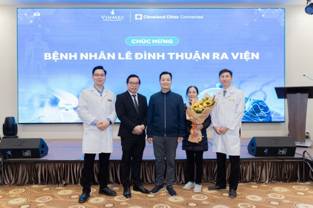
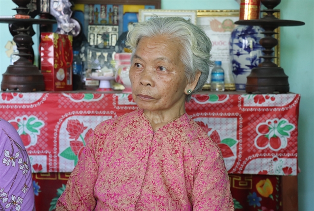
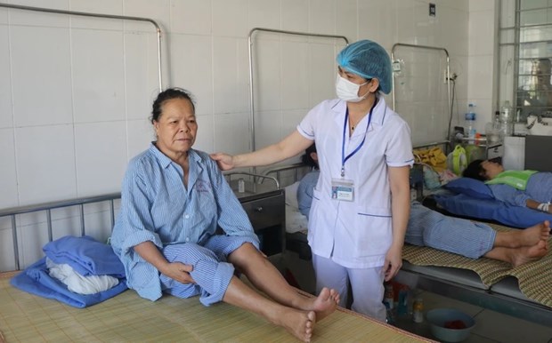
| Patient Thuận is discharged from the hospital on January 22. — Photo courtesy of the hospital |
HÀ NỘI — Vinmec International General Hospital on January 22 announced its doctors simultaneously replaced both the pelvis and part of the femur in one operation to treat a patient with an extremely rare type of bone cancer.
The surgery used new 3D-printed artificial bones designed by a team of Vietnamese doctors and engineers and tested through nearly 100 simulation situations to achieve the highest optimisation, helping to save lives and speed up recovery time for patients.
Patient Lê Đình Thuận, 63, living in Thanh Hóa Province, has very rare bone cancer of the pelvis. His metastatic cancer has invaded the entire structure surrounding the hip joint, including the pelvis, joint capsule and upper femur.
Because it is a complicated disease, many hospitals previously prescribed surgery to remove one side of the pelvis to save the patient's life. However, Thuận has repeatedly refused treatment due to the risk of physical disability and low survival rate after surgery.
When receiving the patient, doctors at Vinmec Orthopedics & Sports Medicine Centre determined that surgery was needed to remove the malignant tissue and reshape the pelvis and femur bones to help the patient move and walk.
“The patient needs surgery as soon as possible to prevent the tumour from progressing and becoming invasive, while ordering from foreign manufacturers takes at least two months. The only way to perform accurate and safe surgery is for a team of surgeons to design the artificial bone implant for the patient,” said Prof. Trần Trung Dũng, Director of Vinmec Orthopedics & Sports Medicine Centre.
“However, the most difficult problem here is choosing the type of material and how to graft artificial bone to recreate the shape and function of the pelvis after it is cut,” said Prof. Dũng.
According to Prof. Dũng, in the world, there have been several cases of 3D pelvic replacements reported. However, to date, there has been no recorded case of simultaneous replacement of the pelvis and upper half of the femur to treat cancer. Other bone shaping solutions, such as using screws and Polyetheretherketone (PEEK) implants, cannot create a biological bond with the pelvis or are not strong enough to withstand body gravity, making it difficult to restore the ability to sit and stand or walk after the surgery.
After more than two weeks of racing against time, continuously testing nearly 100 prototypes of different shapes and structures with situations simulating the load of daily movements of the body, the surgical and engineering teams came up with the most optimal design for the case.
Doctors used a structure that simulates the morphology of the pelvis with a hollow honeycomb shape so that the weight of the entire artificial bone is light, less than half the volume of real bone. The surface in contact with the healed bone is rough with micro-holes to stimulate bone cells to grow onto the artificial bone surface over time to increase stability after surgery.
The material is made from biocompatible medical Titanium alloy. After 3D printing and heating, it is capable of withstanding more than 10 times the load of real bone while still ensuring the same elasticity and durability when compared to the bone tissue of young adults.
The artificial pelvis was also calculated to create fixed positions, helping doctors restore all attachment points of the group of 14 muscle tendons around the pelvis-hip joint.
The sample structure is certified for its mechanical properties to ensure load bearing by the Institute of Materials Science and Engineering, Polytechnic University and the Industrial Testing and Inspection Centre, Institute of Energy Mechanics and Geological Mines.
After the verification process, the pelvic design was sent to Germany for production using a 3D-printing system following European CE medical implant standards.
|
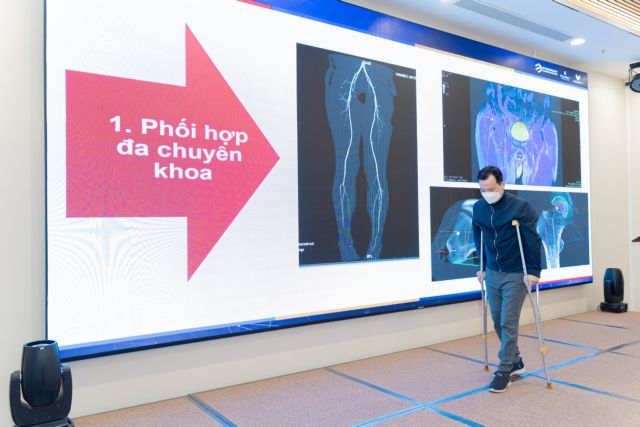
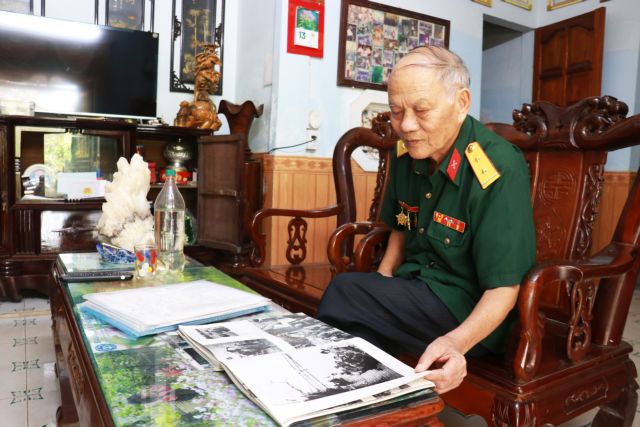
| Patient Thuận could practice walking alone with crutches 10 days after surgery. — Photo courtesy of the hospital |
For the femur, the surgical team used a modular designed titanium artificial femur that has been routinely deployed at Vinmec Hospital for the past three years. The process of manufacturing and importing product samples from abroad only takes one week, much less than the minimum time of two months if ordering from abroad. Therefore, the surgery was promptly performed at the end of December 2023, only about one month after the patient was hospitalised.
The surgery was performed successfully after eight hours without any complications with the participation of many specialties, including vascular surgery, gastroenterology, urology and blood vessel intervention.
The artificial bone of both the pelvis and femur is placed tightly, connecting precisely with the patient's healthy bone. All muscle-tendon attachment points and nerve blood vessels peeled off the bone tumour and were then safely sewn back into the titanium artificial bone graft.
Two days after the surgery, patient Thuận could sit up on his own. After 10 days, he could practice walking alone with crutches for a distance of up to 50 metres without any difficulty. The recovery time, in this case, has been shortened to only one-third compared to some domestic and international reports on artificial pelvis replacement.
Thuận was discharged from the hospital on January 22.
The success of the special surgery has opened up more opportunities for bone cancer patients in Việt Nam, while also creating a solid foundation for Vinmec and VinUni's 3D-printed bone implant product project designed specifically for Vietnamese patients. — VNS
Society
 Society
 Society
 Society
 Society
 Society
 Society
 resize.png) Society
Society
 Society
 Society
 Society
 Society
 Society
 Society
 Society