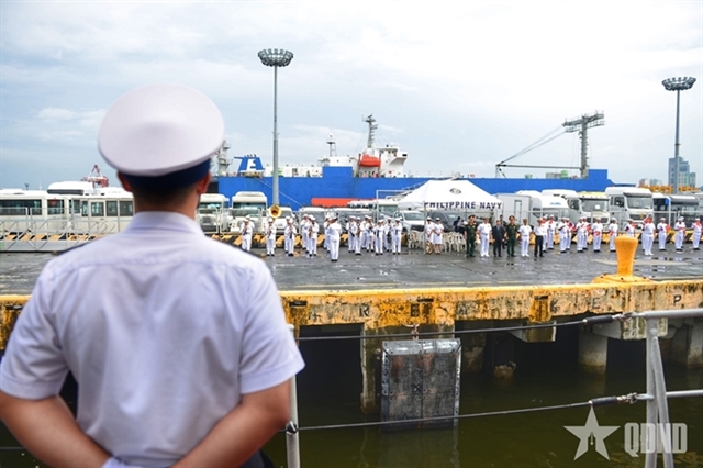
 Society

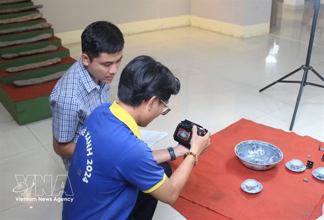
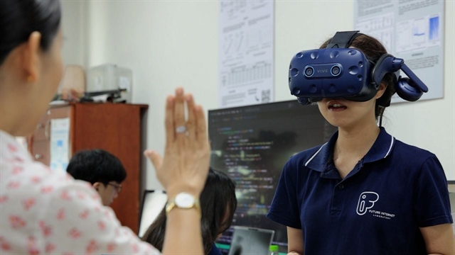
Virtual museums bring Lâm Đồng's heritage into digital age
1.
 |
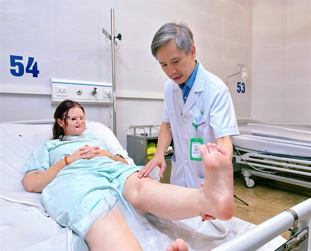
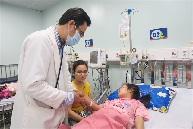
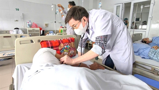
| Dr. Trần Lương Anh (centre), head of the Neurosurgery and Spine Surgery Department at FV Hospital, performs the surgery with his team. — Photo courtesy of the hospital |
HCM CITY — FV Hospital in HCM City has successfully removed a brain tumor measuring over 6cm in diametre by combining vascular embolisation techniques with an advanced neuro-navigation system, and ultrasonic tumor dissection technology.
The tumor was meticulously dissected and removed during a four-hour operation without damaging surrounding brain tissue. Intraoperative blood loss was minimal, and the patient made a remarkable recovery just two hours after surgery.
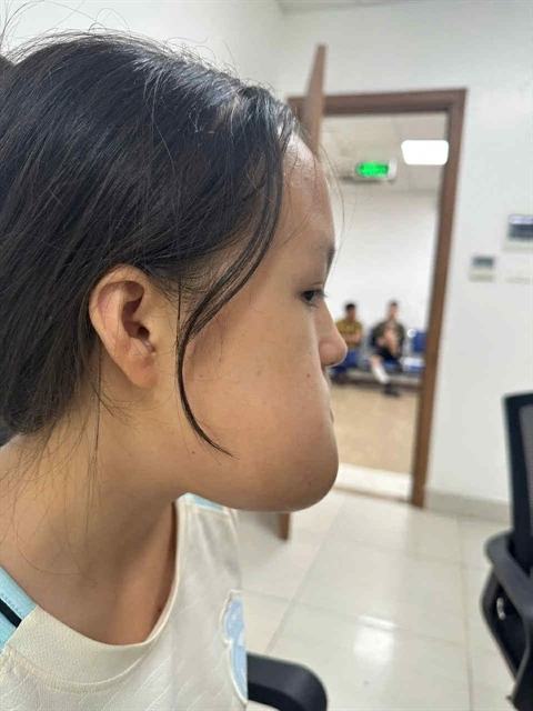
The patient, Trịnh Nhật Khánh (45, residing in HCM City), had occasionally experienced mild headaches and decided to undergo stroke screening at FV Hospital. An MRI scan revealed a large meningioma measuring over 6 cm in diameter—about the size of an orange—compressing nearly one-quarter of her brain.
According to Dr. Trần Lương Anh, head of the Neurosurgery and Spine Surgery Department at FV Hospital, the tumor had grown over a long period, allowing the skull to adapt and making it difficult to detect. Though the patient experienced some symptoms, the tumor had fortunately not affected higher neurological functions. Still, timely surgery was essential, as further growth could lead to severe complications such as seizures or become life-threatening.
Following clinical consultation, doctors opted for surgery despite several risks. The tumor’s size and firmness made removal challenging and increased the potential for damage to surrounding brain tissue. Moreover, its dense vascular supply posed a high risk of heavy blood loss during the surgery.
To reduce intraoperative bleeding and preserve critical blood vessels, the surgical team performed a pre-surgical embolisation procedure one day prior to surgery to block the tumor’s blood supply. The following day, the operation, led by Anh, was conducted with the support of the neuro-navigation system, which allowed precise identification of tumor margins and helped optimise the surgical pathway. This minimised the incision size, shortened the surgery time, and reduced surgical risks. The system also enabled accurate mapping and protection of nearby blood vessels.
The ultrasonic tumor dissection device also played a key role in addressing the tumor’s hardness. According to Anh, for large and firm tumors, manual removal can endanger adjacent brain tissue. The ultrasonic device enables tumor removal with minimal movement, reducing impact on brain tissue and protecting adjacent blood vessels.
The multi-modality surgery was completed successfully in four hours. The tumor was fully removed with minimal bleeding, no damage to surrounding tissue, and a notably fast recovery. Four days post-surgery, the patient had regained approximately 90 per cent of her normal health. Pathological results confirmed the tumor was benign. She is expected to be discharged soon and return to daily life.— VNS
Society
 Society
 Society
 Society
 Society
 Society
 Society
 Society
 Society
 Society
 Society
 Society
 Society
 Society
 Society